How the brain works · 01
Neuro-Anatomy
Structure and location in the brain. Specific sites in your brain are responsible for specific functions, so training different locations gives you different outcomes.
Vision, hearing and other senses
You don’t see with your eyes. You see with your brain.
The very back of your brain, the occipital lobes, contains the primary and secondary visual cortex. These areas are entirely responsible for creating and processing vision from signals that come from your eyes.
Of course you need your eyes: they take in light through the lens and focus it onto the retina, where rods and cones convert light into electrical activity. That activity travels through the optic nerve (the 2nd cranial nerve) to the visual cortex, where vision is actually created. Through connections with other parts of your brain, you then interpret what you see and decide how to react.
The same is true of all your senses. Your ears don’t create hearing: sound moves your eardrum, tiny inner-ear bones help create electrical signals, and those travel through the auditory nerve (the 8th cranial nerve) to the auditory cortex in your temporal lobe. Smell works the same way through the olfactory nerve (1st cranial nerve). As you might guess, dogs devote a much larger portion of their brains to smell than humans do.
Vision
Occipital lobeRetina → Optic nerve (CN II) → Visual cortex
Hearing
Temporal lobeEardrum → Auditory nerve (CN VIII) → Auditory cortex
Smell
Olfactory cortexNose → Olfactory nerve (CN I) → Olfactory cortex
Touch & pain
Somatosensory cortexNerve endings → Spinal cord → Pain Network
Body sensations and pain
The Pain Network
Sensation from your entire body travels through your nerves and spinal cord directly to your brain, where sensations, including pain, are created and processed.
Key locations
The most important locations for pain form a complex web of connections that create and process pain and suffering.
Thalamus
Somatosensory cortex (SSC)
Insular cortex
Anterior cingulate cortex (ACC)
Three pain pathways
Three distinct pathways send signals to different parts of the Pain Network.
-
01
Primary pain pathway
Carries the basic signal of where and how intense a sensation is.
-
02
Suffering pain pathway
Adds the emotional, distressing quality of pain.
-
03
Pain inhibition pathway
Your brain’s built-in system for turning pain down.
Too much activity in the suffering pathway and too little in the inhibition pathway almost always means chronic pain, whether chronic migraines, face pain, neck, shoulder or back pain, IBS, pelvic pain, or extremity pain such as CRPS.
Location of pain in your body
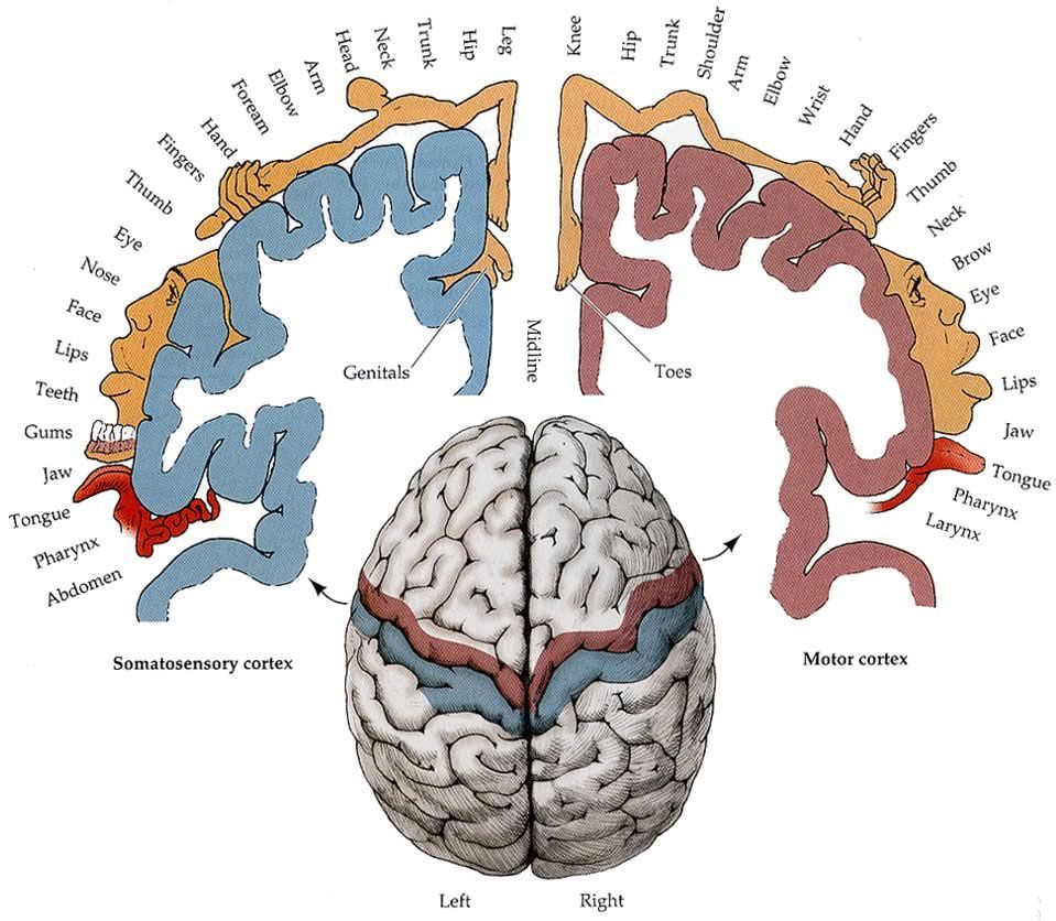
The homunculus: a map of you, inside your brain
Where you feel chronic pain depends on exactly which part of your Pain Network has abnormal electrophysiological (EP) activity. Your brain has every part of your body mapped to specific locations.
If a specific part of the SSC is stimulated, for example the face area of the right SSC, you feel that sensation in the matching body part (in this example, the left side of your face). Neurosurgeons sometimes use this stimulation during brain surgery, such as removing a tumor, to avoid areas that control movement and sensation.
The very top of the SSC and motor cortex controls your feet; the bottom controls your face, with every other body part mapped in between. Draw the body parts in proportion to the brain area devoted to them and you get a funny-looking “little human,” the homunculus.
This explains phantom pain: someone can feel severe pain in a foot that was amputated years ago, because the part of the brain responsible for that foot is still active. Phantom pain can only be explained by pain being created in the brain.
All pain is created in the brain
If you step on a nail, the nail doesn’t create the pain; it starts the process. Nerve endings send electrical signals up your leg and spinal cord to your Pain Network, where the decision for pain is made.
Because that’s true, a brain map can show the abnormal activity in your Pain Network, and we can use that exact information to turn its intensity down, so pain becomes more tolerable or goes away completely.
How we treat chronic painReady to see what your brain map reveals?
Book a consultation in our Nashville office or by video from anywhere in the world. Our team will review your symptoms and recommend a personalized plan.